What is calcific tendonitis?
 What is calcific tendonitis?
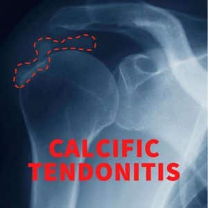
Calcific tendonitis of the shoulder is also referred to as calcific tendinitis or calcific tendinopathy is a common condition that can affect the rotator cuff tendons of the shoulder. The exact cause of the condition is not fully understood but we know it is more common in females, tends to affect those in the middle age, and is associated with endocrine disorders such as hypothyroidism.
How does calcium end up in my shoulder tendon?
While the exact trigger for calcium deposition is not fully understood, it is believed that the process starts with tendon degeneration. In some individuals, tendon cells are transformed to cartilage forming cells and they deposit calcium hydroxyapatite crystals into the tendon.
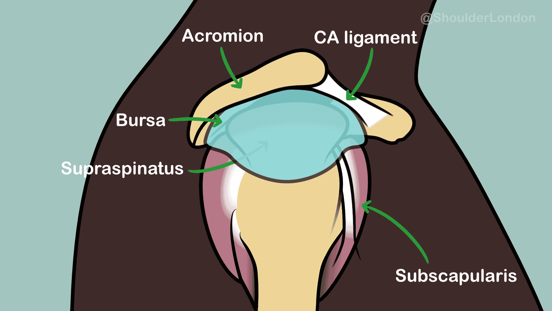
Side view of normal anatomy of the shoulder. (Click to enlarge)
In the shoulder the rotator cuff tendon most commonly affected is the supraspinatus tendon at the top of the shoulder.
Calcific tendonitis of the shoulder undergoes several stages:
- Pre-calcific stage (no pain)
- Formative stage (pain may by absent or mild)
- Resting stage (pain may be absent or mild)
- Resorptive phase (acute and often severe pain)
- Post-calcific stage (pain may be absent or mild)
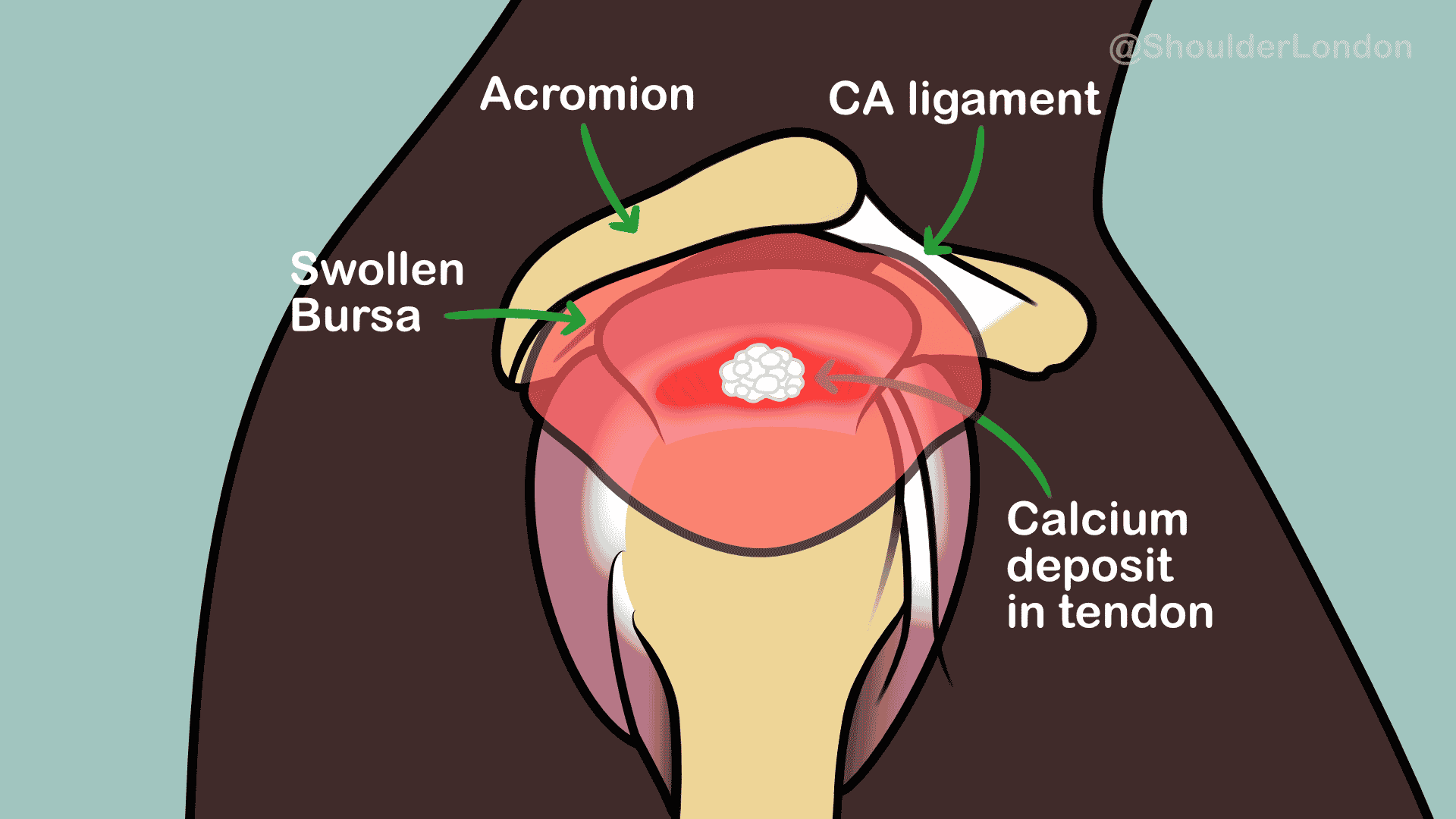
Diagram showing calcium in supraspinatus tendion with bursitis. (Click to enlarge)
Calcium forms in the tendon during the formative phase and usually gets resorbed spontaneously in the resorptive phase due to an inflammatory process. In the post-calcific phase, healing usually occurs in the tendon at the site of calcification.
What are the symptoms of calcific tendonitis?
During the formative and resting phase, the patient may have no symptoms or may have some mild chronic pain in the shoulder. During the resorptive phase the inflammatory response to the calcium can trigger a sudden onset of severe acute pain. Patients often describe waking up in severe pain without a history of injury. The pain may limit the ability to move.
How is calcific tendonitis diagnosed?
Calcium is well seen on x-ray so usually x-rays of the shoulder are all that is needed to make the diagnosis. Occasionally we may use MRI to assess the rotator cuff integrity in severe cases to ensure there isn’t a tear in the tendon. In the formative and resting phases, the calcium usually takes on a well-defined appearance. In the resorptive phase, the calcium can look patchy or fluffy on x-ray.
Can calcific tendonitis go away on its own?
Not all patients with calcific deposits in their rotator cuff tendons have pain or symptoms, we occasionally see the calcium on x-rays taken for other issues. The calcium deposited in the tendon gets resorbed naturally so the condition can resolve on its own. Unfortunately, some patients may experience different lengths of phases or may get stuck in a particular phase.
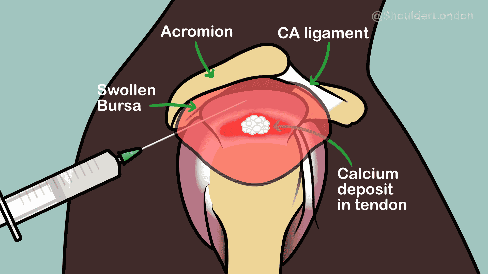
Steroid injection into the subacromial bursa over calcific deposit. (Click to enlarge)
Calcific tedonistis treatment, what are the options?
Calcific tendonitis treatment options depend on the phase of the condition and the symptom severity. In the formative and resting phases, over the counter pain killers may be all that are needed to manage pain. Sometimes a subacromial space steroid injection may be needed if there are signs of subacromial impingement. In the resorptive phase, when there is acute severe pain, stronger pain killers may be necessary. Physiotherapy is useful to prevent the patient from becoming stiff. Steroid injections in this phase should be considered carefully as this may switch off the inflammation that is required to remove the calcium naturally.
Other non-operative calcific tendonitis treatments include ultrasound guided barbotage, where the calcium is
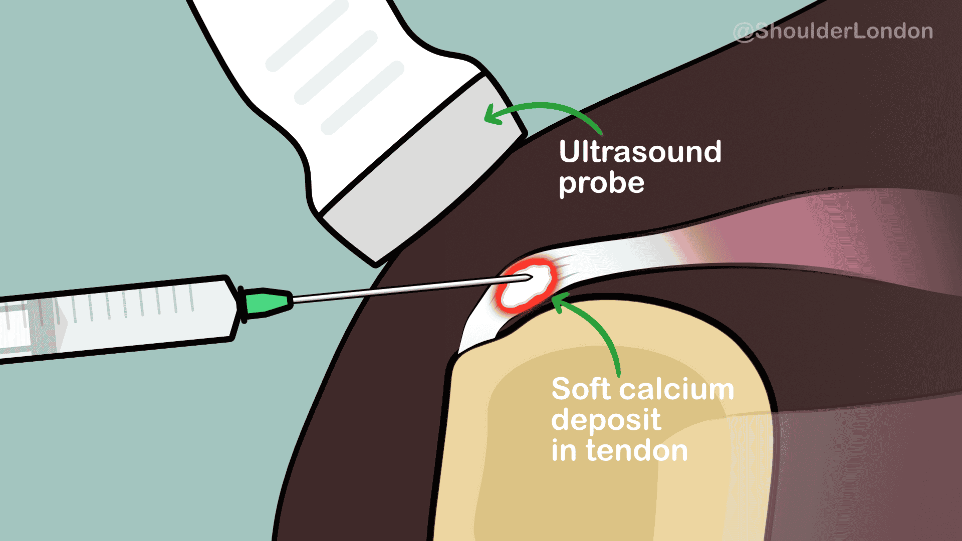
Ultrasound guided barbotage. (Click to enlarge)
flushed out of the tendon through needles using ultrasound guidance. Extra-corporeal shockwave therapy (ESWT) is another treatment option available where pressure waves generated by a machine are directed toward the calcification of the tendons. This breaks up the calcium and triggers an inflammatory response which helps to resorb it.
If non-operative calcific tendonitis treatment fails, then surgical excision of the calcium can be performed through key-hole surgery (arthroscopy). In the formative or resting phase, the calcium is firm and has to be scooped out of the tendon, while in the resorptive phase the calcium is more toothpaste like and can be squeezed out of slits made in the tendon.
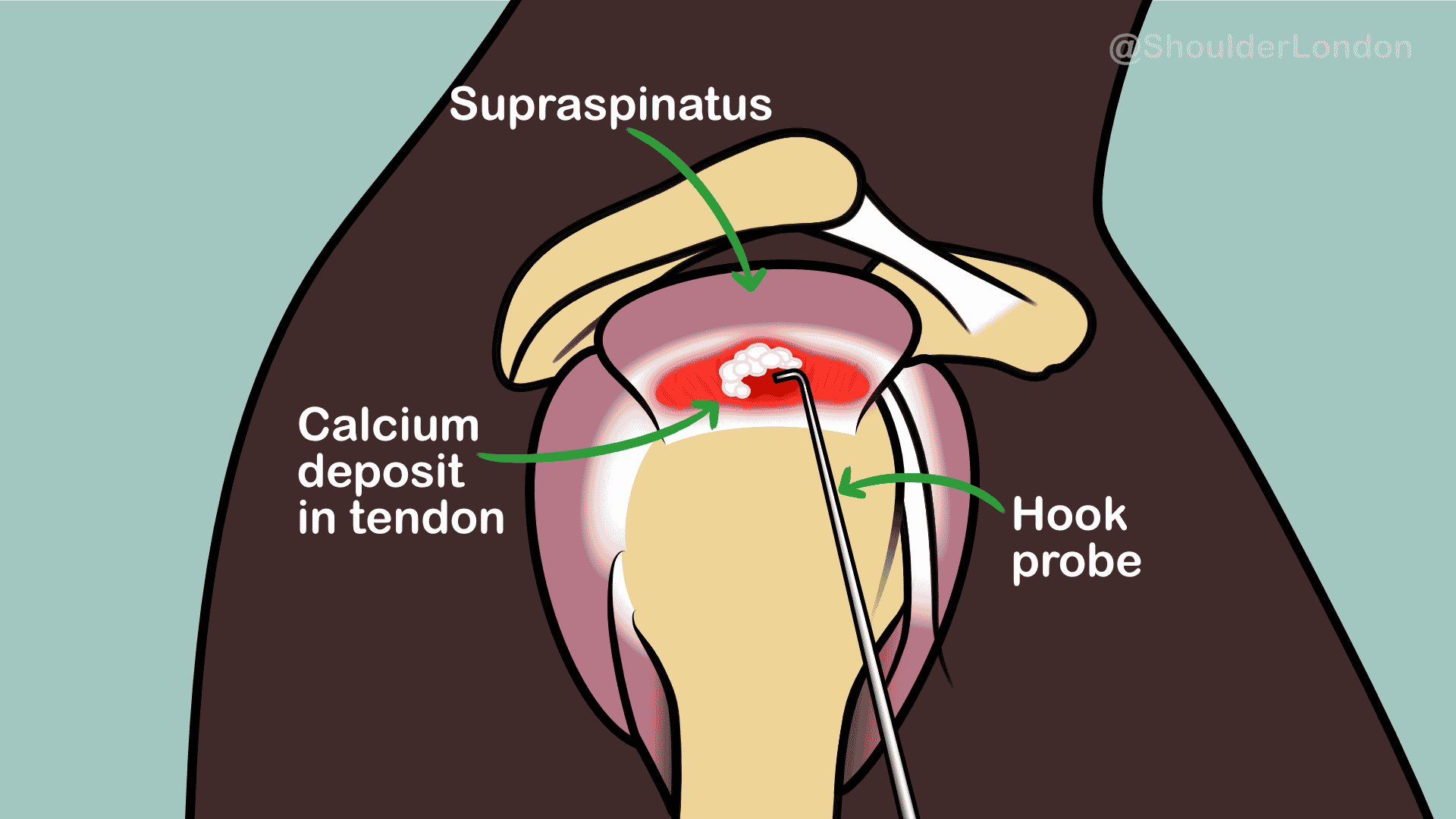
Arthroscopic excision of calcium deposit. (Click to enlarge)
In chronic cases the calcium may be difficult to locate, and excise, and other causes of pain need to be excluded and treated.
If you think you have the symptoms of calcific tendonitis of the shoulder contact us to arrange a consultation.
Nick Ferran @ Shoulder & Elbow London Ltd
Clinics in:
Chiswick – Harrow – St. Johns Wood
As an Amazon Associate, I earn from qualifying purchases.